Femoral vein and artery
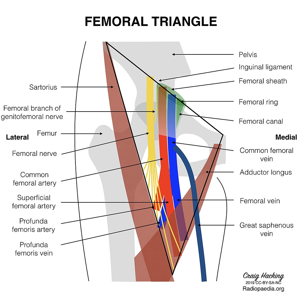
Femoral triangle
3D Tour of the Femoral Triangle by About Medicine CC BY-SA 4.0 via Wikimedia Commons
Borders
| Direction | Relation |
|---|---|
| Superior | inguinal ligament |
| Lateral | sartorius muscle |
| Medial | adductor longus muscle |
| Deep | pectineus, ilacus and psoas muscles |
| Superficial | see surface anatomy |
Contents
Mnemonic - NAVEL
The contents of the femoral triangle can be remembered by the mnemonic NAVEL (nerve, artery, vein, empty space, lymphatics).
NAVEL describes the structures in order from lateral to medial at the superior aspect of the triangle.
- Femoral nerve and its muscular branches
- Femoral sheath
- Femoral artery and vein
- The relationship of the femoral vein and artery changes as the vessels descend in the femoral triangle
- Proximal/superior femoral triangle
- The femoral vein is medial to the femoral artery
- Distal/inferior femoral triangle
- As the femoral vein descends in the femoral triangle, it moves posterolaterally to the artery (behind and around it), so that it is lateral to the femoral artery in the distal femoral triangle.
- Femoral canal
- contains empty space, lymphatic vessels and the lacunar lymph node
- the lymphatic vessels drain into the external iliac nodes
- Femoral artery and vein
Surface anatomy
- The femoral artery is found at the midinguinal point (midpoint between pubic symphysis and ASIS)
- The femoral vein is 0.5-1cm medial to this
- Layers traversed from skin to vessel:
- skin, subcutaneous fat, superficial fascia, fascia lata, femoral sheath, vessel
Origin and course of the femoral vein
- Also known as the common femoral vein
- Drains from popliteal vein
- Travels within femoral sheath
- Drains into external iliac vein at inguinal ligament → common iliac → IVC
- Tributaries
- great saphenous vein
- enters the femoral sheath within femoral triangle via fossa ovalis (medial aspect of thigh)
- it drains superficial the superficial lower limb veins
- deep femoral vein - from deep thigh
- medial and lateral circumflex veins - from femoral head
- great saphenous vein
Origin and course of the femoral artery
Terminology
'Femoral artery' refers to both the common femoral (CFA) and superficial femoral (SFA) arteries.
Distinguishing between the two is considered superfluous and misleading by Terminologia Anatomica, the international standard for human anatomical terminology, as the SFA is not actually superficial.
- The femoral artery is a continuation of external iliac artery at the level of the inguinal ligament
- Enters thigh posterior to inguinal ligament
- Variable length up to 8cm, and up to 1cm wide
- First 4cm is enclosed within femoral sheath with femoral vein
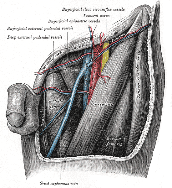
- Gives rise to:
- 3 superficial arteries
- superficial epigastric → abdo wall
- superficial circumflex iliac
- superficial external pudendal
- 2 deep arteries
- deep external pudendal → pelvis/pelvic wall
- deep femoral artery (aka profunda femoris) → deep tissues
- gives rise to medial and lateral circumflex arteries which wrap around the femur
- 3 superficial arteries
- Leaves femoral triangle posterior to sartorius in adductor hiatus
- Continues into lower leg as popliteal artery

Human leg bones labeled by Jecowa CC BY 3.0 via Wikimedia Commons
ON THIS PAGE