Bacterial classification
Outline the process of Gram staining
crystal Violet stain
Iodine
Solvent wash (Alcohol/Acetone)
Safranin counterstain
Gram positive bacteria have a thick peptidoglycan cell wall, while gram negative bacteria have a thin peptidoglycan wall with an outer lipopolysaccharide layer. Gram staining leverages the fact that stains wash out of thin-walled bacteria more easily, to apply a different colour to gram positive and negative bacteria.
Crystal violet stain
Stains both gram positive and gram negative bacteria purple.
Crystal violet dissociates to a positively charged ion which binds to negatively charged components of bacterial cells, staining them purple.
Lugol's iodine solution
A mix of iodine and potassium iodide, Lugol's iodine is used as a trapping agent. The negatively charged iodide forms a large, stable complex with the crystal violet stain, preventing its removal.
Solvent wash
A solvent wash (typically alcohol or acetone) destroys the outer lipopolysaccharide layer of gram negative bacteria, exposing the thin peptidoglycan layer.
This allows the crystal violet stain to wash out of gram negative bacteria through the thin peptidoglycan wall, leaving them colourless.
If the wash is left on for too long, it will wash the crystal violet out of gram positive bacteria as well.
Safranin counterstain
A counterstain that then stains Gram negative bacteria pink. Safranin is positively charged too, just like crystal violet. It is also taken up by gram positive bacteria but cannot be seen against the much darker crystal violet stain.
At the end of this process, gram positive bacteria remain purple while gram negative bacteria are stained pink.
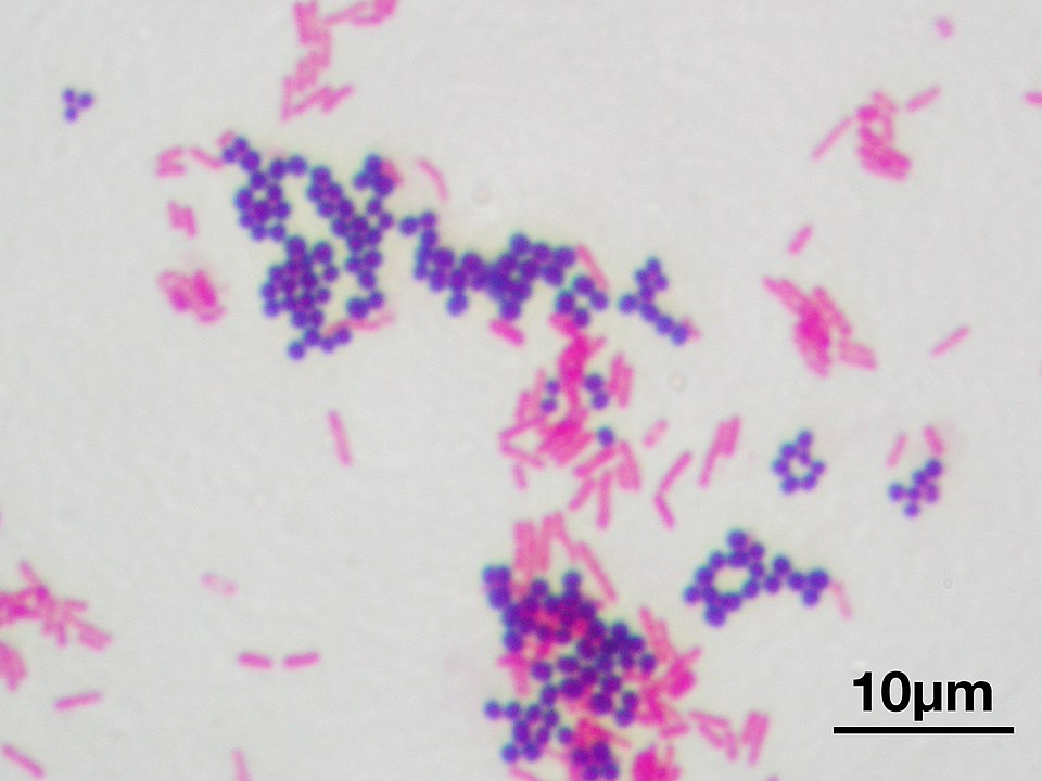
A culture of gram-positive Staphylococcus aureus, stained purple, with gram-negative Escherichia coli, stained pink.
By Y tambe CC-BY-SA 3.0 via Wikimedia Commons
Classify bacteria with examples
Gram positive bacteria
Classification | Examples | ||
|---|---|---|---|
Cocci | Staphylococcus | coagulase +ve | S. aureus |
coagulase -ve | S. epidermis | ||
Streptococcus | α-haemolytic | S. pneumoniae S. viridans | |
β-haemolytic | GAS - S. pyogenes GBS - S. agalactiae | ||
γ-haemolytic | S. bovis | ||
Enterococcus | E. faecium E. faecalis | ||
anaerobes | Peptococcus spp. Peptostreptococcus spp. | ||
Bacilli | aerobes | *Bacillus spp. (B. cereus, B. antdracus) | |
Lysteria monocytogenes | |||
anaerobes | *Clostridium spp. (C. difficile, C. botulinum, C. tetani) | ||
*considered gram-variable due to an increasing number of gram negative cells as the culture ages | |||
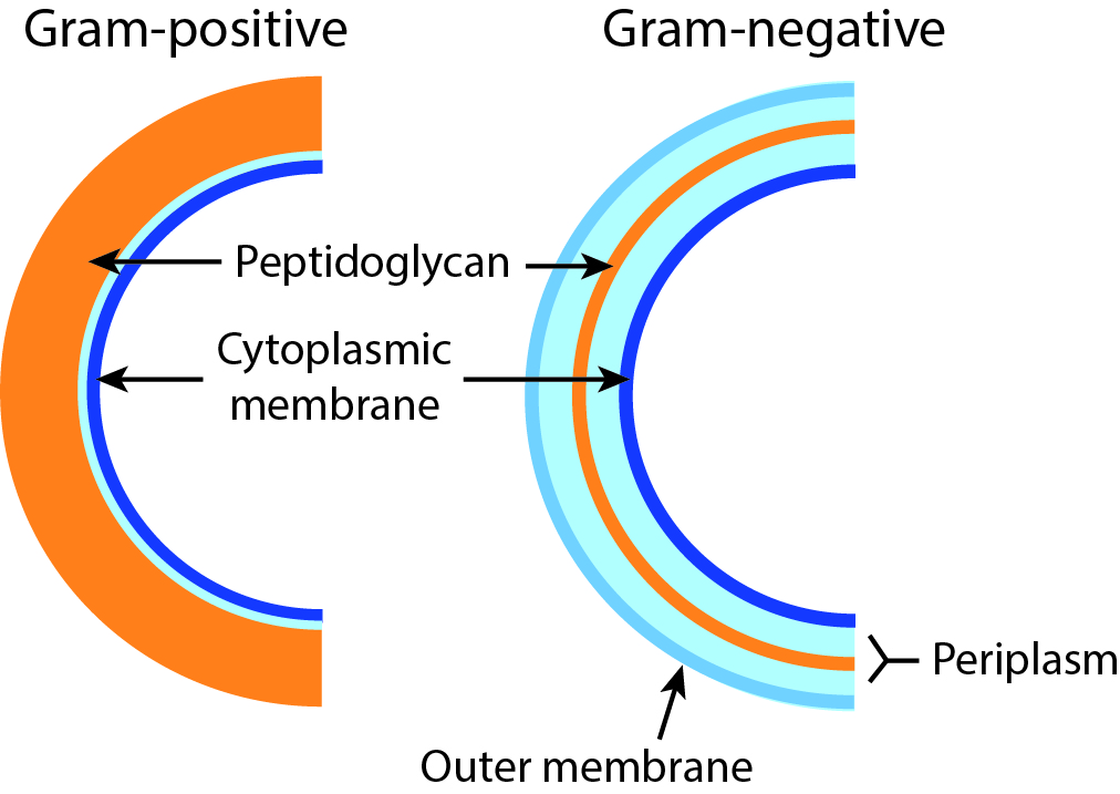
Gram positive bacteria have a thick peptidoglycan wall, while gram negative bacteria have a think peptidoglycan wall with an outer lipopolysaccharide membrane.
By Microbialmatt CC-BY-SA 4.0
Gram negative bacteria
Classification | Examples | ||
|---|---|---|---|
Cocci | aerobes | Neisseria spp. N. gonorrhoea, N. meningitidis | |
Moraxella catarrhalis | |||
Bacilli | short | Haemophilus influenza | |
Bordatella pertussis | |||
Legionella pneumophilia | |||
long | facultative anaerobes | Proteus mirabella | |
Salmonella spp. S. typhi, S. enterica | |||
Shigella spp. S. dysenteriae | |||
Yersinia pestis | |||
obligate anaerobes | Pseudomonas aeroginosa | ||
curved or spiral | microaerophilic | Helicobacter pylori | |
Campylobacter jejuni | |||

A closer look at the cell wall of gram negative bacteria
By Jeff Dahl CC-BY-SA 4.0 via Wikimedia Commons
Gram indeterminate bacteria
So called because they Gram stain poorly.
Examples:
- Mycoplasma spp. (e.g. M. pneumoniae) — pleomorphic bacteria that lack a cell wall
- Mycobacterium spp. (e.g. M. tuberculosis) — bacilli that resist decolourisation by acids, hence known as acid-fast bacilli