The parasympathetic nervous system
Define the parasympathetic nervous system
Part of the autonomic nervous system that controls various unconscious functions, mostly via control of visceral organs. May be considered a physiological brake.
Outline the anatomy of the vagus nerve
Functional pathways
The vagus nerve (cranial nerve X) is a long, important nerve that has many fibre types which serve various parasympathetic and somatic functions:
- visceral motor fibres
- produce secretory and motor effects in the heart, vasculature, bronchi, and GI tract
- visceral sensory fibres - aortic baroreceptors and chemoreceptors - dura - heart, respiratory tract and GI tract - epiglottis (taste)
- somatic sensory fibres, supplying the external auditor meatus
- somatic motor fibres, supplying the skeletal muscles of larynx/pharynx/upper oesophagus
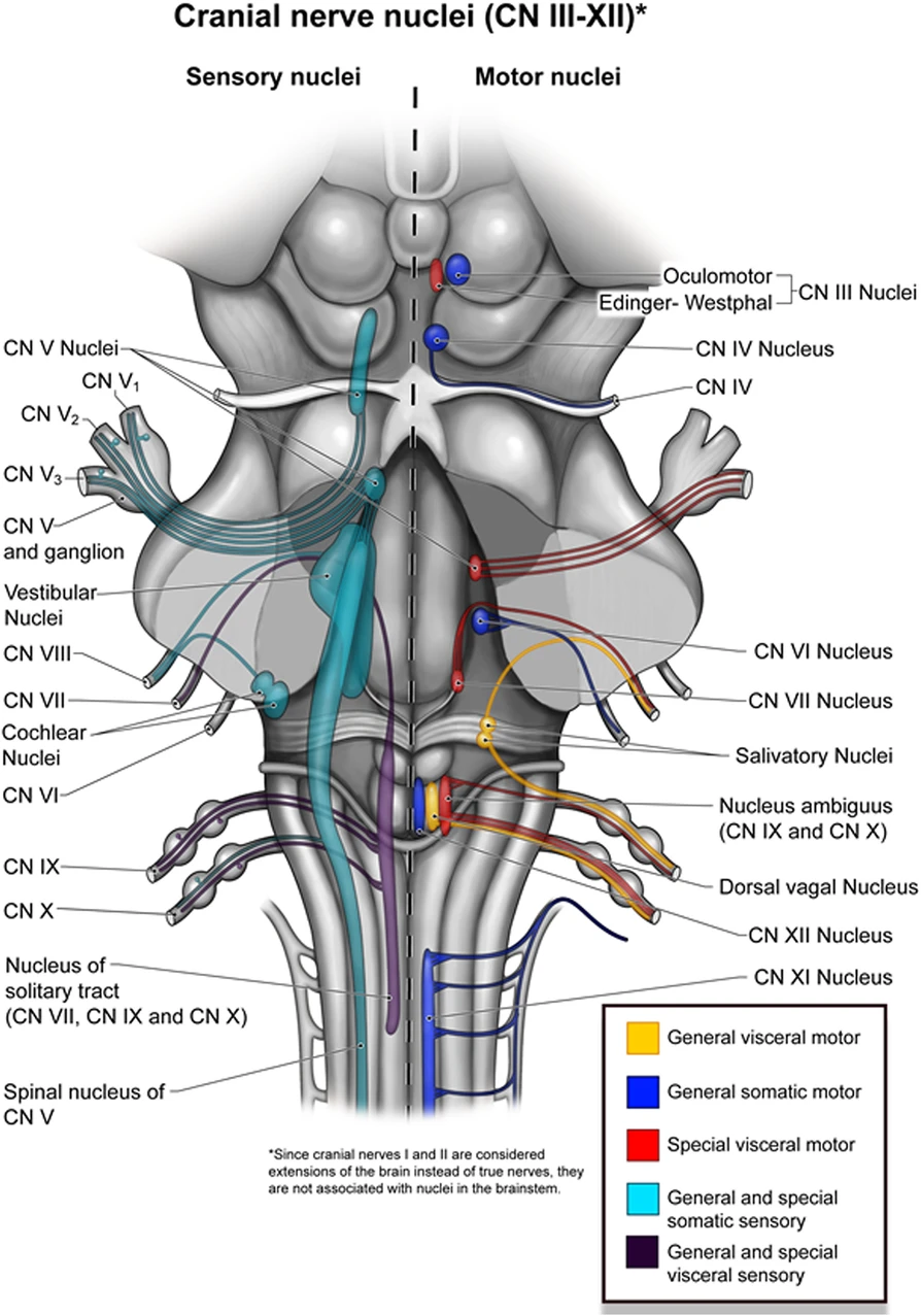
Origin
The vagus nerve originates from 4 nuclei in the medulla oblongata
- Dorsal vagal nucleus - carres mixed visceral sensory/motor fibres
- Nucleus ambiguus (NA) - carries motor fibres to the pharynx/larynx (known as ‘branchial’ motor neurones)
- Nucleus tractus solitarius (NTS) - carries sensory fibres including taste
- Spinal trigeminal nucleus - a minor contribution to the vagus nerve, carrying somatic sensory fibres
By Montoya, S., Portanova, A. & Bhatt, A.A, licensed under CC-BY 4.0
Course
Head and neck
- Meningeal, Auricular, Pharyngeal, SLN, cardiac
Chest
- Cardiac, RLN, Oesophageal, Pulmonary
Abdomen
- vagal Trunks, Hepatic, Coeliac, Gastric
Head
Exits the cranium via the jugular foramen
There are two sensory ganglia at this level:
- superior ganglion of the vagus nerve (aka jugular ganglion)
- gives off the meningeal branch → dura
- inferior ganglion of the vagus nerve (aka nodose ganglion)
- gives off the auricular branch → external auditory meatus
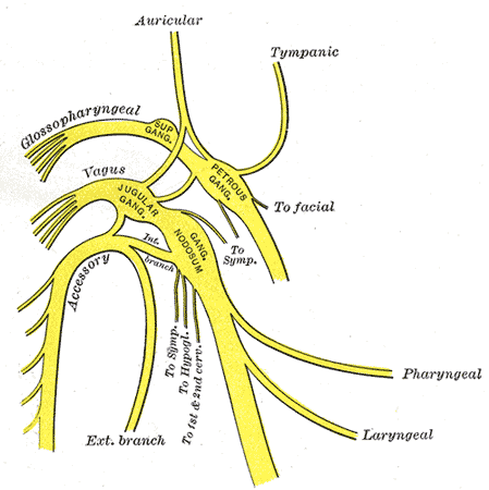
Cranial ganglia of the vagus nerve.
Neck
Descends in carotid sheath between internal jugular vein and carotid artery, lateral to trachea.
Branches
- pharyngeal branch → pharyngeal plexus
- superior laryngeal nerve (SLN) → larynx (motor and sensory)
- cardiac branches → superficial and deep cardiac plexuses

Superior and recurrent laryngeal nerves.
By Jkwchui, licensed under CC-BY-SA 3.0
Chest
Enters thorax anterior to both subclavian arteries.
Branches
- cardiac branches → superficial and deep cardiac plexuses
- recurrent laryngeal nerves (RLN) → neck → larynx - L) RLN passes under the aortic arch
- R) RLN passes under the R) suvbclavian artery
- oesophageal plexus → oesophagus
- pulmonary branches - anterior pulmonary branches → anterior pumonary plexus
- posterior pulmonary branches → posterior pulmonary plexus
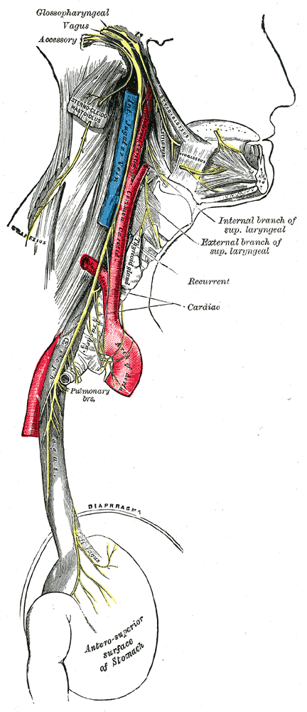
Branches and plexuses of the vagus nerve.
Abdomen
Enters abdomen through oesophageal hiatus in diaphragm at the level of T10 - L) vagus → anterior vagal trunk - R) vagus → posterior vagal trunk
Branches - gastric branches → stomach - coeliac branches → coeliac plexus and superior mesenteric plexus → renal/adrenal/GI - hepatic branches → hepatic plexus
Imagine you are in a tiny car driving inferiorly down the oesophagus towards the stomach.
When you reach the stomach, you are forced to turn right because that is the direction the stomach takes.
As you turn the steering wheel, your left hand moves from the left side of the wheel to the anterior side of the wheel, while your right hand moves to the posterior side of the wheel.
The vagus nerves takes the same path as it descends down the oesophagus. When it reaches the stomach, it too turns right. The left vagus nerve ends up anterior, forming the anterior trunk. The right vagus nerve ends up posterior, forming the posterior vagus trunk.
Outline the non-vagal components of the parasympathetic nervous system
Cranial
CN III (oculomotor nerve)
- the parasympathetic nucleus of CN III is known as the Edinger-Westphal nucleus
- it supplies the ciliary ganglion → pupil constriction and accomodation
CN VII (facial nerve)
- supplies two ganglia:
- pterygopalatine ganglion → lacrimal gland → lacrimation
- submandibular ganglion → submandibular/submental salivary glands → salivation
CN IX (glossopharyngeal nerve)
- supplies the otic ganglion → parotid gland → salivation
- also receives signals from the carotid baroreceptors and chemoreceptors
Sacral
Pelvic splanchnic nerves originate from S2-4. Often described as ‘the mechanism for emptying'.
- gastrointestinal effects
- anal sphincter relaxation
- stimulates distal colon (from splenic flexure) and rectum peristalsis
- genitourinary effects
- detrusor contraction
- bladder sphincter relaxation
- erection
The effect of the autonomic nervous system on male genitalia is summarised by the phrase 'point and shoot'.
-
PNS controls the Pointing, or erection.
-
SNS controls the Shooting, or ejaculation (via contraction of seminal vesicles)
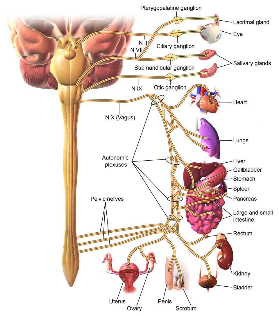
Innervation of the parasympathetic nervous system
By Blausen Medical, licensed under CC-BY 3.0
Outline the nerve fibre types and synaptic physiology
Nerve fibres
Preganglionic | Postganglionic | |
|---|---|---|
Length | Long | Short |
Fibre type | Myelinated B-fibres | Unmyelinated C-fibres |
Synapse location | Autonomic ganglia close to the target organ | On target organ |
Neurotransmitter released | Releases acetylcholine (ACh) which activates type-2 nicotinic (N2) receptors on postganglionic nerves | Releases ACh which activates muscarinic receptors on target organs |
Receptors and neurotransmitters
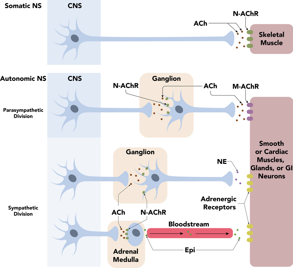
Pre- and post-ganglionic nerve and neurotransmitter types in parasympathetic, sympathetic, and somatic neurones.
By Daniel Walsh and Alan Sved, licensed under CC-BY-SA 4.0
References
Ellis, Feldman, Harrop-Griffiths, Lawson. Anatomy for anaesthetists, 8th edition. Blackwell Publishing. 2004.